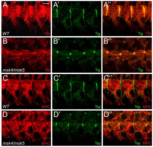
Figure 6. The ECM proteins Tig and Tsp are secreted and localized to muscle attachment sites.
(A-D”) Ventral musculature with anterior to the left and dorsal up. (A-D”) The ventral musculature (red) in stage 16 embryos stained with ECM proteins (green). (A-B”) Tig is enriched at muscle-muscle attachment sites in wild-type embryos (A-A”). The localization and expression of Tig is still present in msk mutant embryos, although the size of the attachment site is reduced (B-B”). (C-D”) Tsp is enriched at muscle-tendon attachment sites in both wild-type (C-C”) and msk mutant embryos (D-D”). As with Tig, there is a reduction in the size of the attachment sites. Scale bar: 20 μm.

