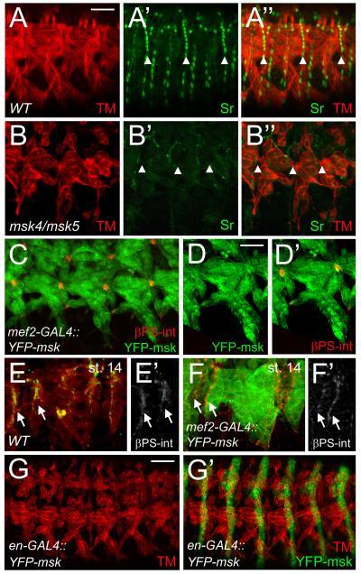
Figure 8. Msk is necessary, but not sufficient for tendon cell induction.
Immunofluorescent stainings. Ventral musculature with anterior to the left and dorsal up. (A-B”) Sr protein is found in all mature tendon cells in stage 16 embryos (A’-A”). A decrease in Msk levels results in a dramatic decrease in Sr protein (B’-B”). Muscle is shown in red. (C-D”) Expression of YFP-Msk in the musculature with mef2-GAL4 results in either a WT muscle pattern (Figure 4H) or muscles that appear pointed at the ends and have smaller attachment sites as visualized by βPS-integrin staining (C-D”). (E-F’) βPS-int is present at the ends of muscles in stage 14 embryos in WT (E, E’) and in muscles that are over-expressing Msk (F, F’). (G-G ’) Expression of YFP-Msk in all epidermal cells under control of the engrailed promoter shows muscles (red) with aberrant muscle attachments. In all examples, anterior is left and dorsal is up. All panels are representative embryos of ventral muscles. Scale bars: 20 μm in A-C; 10 μm in D-D’; 50 μm in G-G’.

