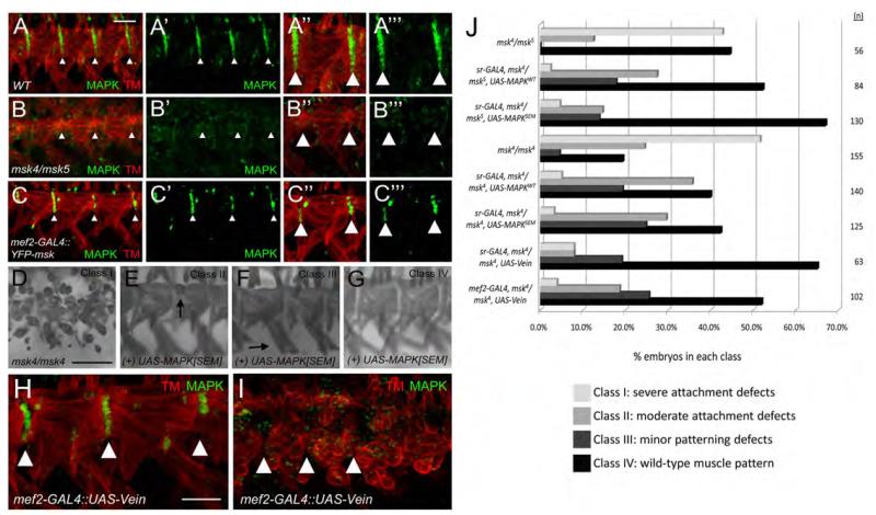
Figure 9. MAPK and Vein act downstream of Msk to mediate muscle-tendon attachment.
(A-C’”) Immunofluorescent stainings of muscle (red) and activated MAPK (green). (A-A’”) In WT embryos, MAPK is present in the tendon cells and extends the length of the ventral muscles at the segment borders. (B-B’”) In msk mutants, activated MAPK cannot be detected at the muscle-tendon attachment sites. (C-C’”) The number of tendon cells that show activated MAPK staining is reduced in embryos that over-express Msk in the musculature. (D-G) MHC staining. (D) Severe attachment defects are observed in msk4/msk4 embryos (Class I). Expression of activated MAPK (MAPKSEM) in the tendon cells using sr-GAL4 results in muscles with mild attachment defects (E; Class II), patterning defects (F; Class III), or complete rescue to a WT muscle pattern (G; Class IV). (H, I) Expression of Vein in the musculature results in smaller attachment sites (compare to Figure 9A-A’”) and reduced MAPK staining (H) or severe attachment defect and a complete loss of activated MAPK at the attachment sites (I). (J) Bar graph that quantitates the rescue of muscle attachment defects in msk4/msk5 or msk4/msk4 mutant embryos upon over-expression of WT MAPK, activated MAPK, or Vein. In all examples, anterior is left and dorsal is up. All panels are representative embryos of ventral muscles. Scale bars: 20 μm in A-G; 10 μm in H-I.

