Figure 4.
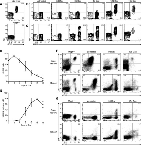
Pax5 restoration triggers leukemia differentiation and regression. (A) Flow cytometry of CD19 and IgM expression in peripheral blood of wild-type (top panel) and untransplanted Rag1−/− (bottom panel) control mice. (B) Flow cytometry of CD19 and IgM expression on mononuclear cells from the peripheral blood of a representative Rag1−/− mouse transplanted with STAT5-CA;Vav-tTA;TRE-GFP-shPax5 leukemia and Dox-treated upon leukemia development as indicated. (C) Time course analysis as shown in B for an independent leukemic recipient mouse. (D) Leukemia burden (proportion of CD19+ cells in the blood) upon Dox treatment (mean ± SEM; n = 3 mice). (E) Proportion of CD19+ cells coexpressing IgM upon Dox treatment (mean ± SEM; n = 3 mice). (F) Flow cytometry of CD19 and CD93 expression on bone marrow cells (top panels) and splenocytes (bottom panels) isolated from representative leukemic Rag1−/− mice at various times during Dox treatment as indicated. The left panels are profiles from untransplanted Rag1−/− control mice, indicating a small population of CD19+ pro-B cells in the bone marrow. (G) Flow cytometry of CD19 and IgM expression as shown in F.

