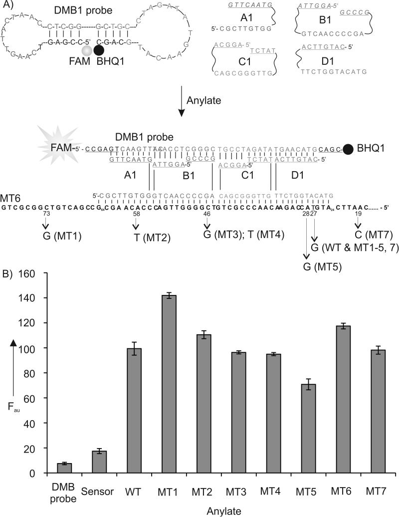
Figure 2.
The structure and performance of Sensor 1. A) Schematic diagram of Sensor 1 in the absence and in the presence of MT6 analyte. DMB-binding arms of adaptor strands A1, B1, C1 and D1 are underlined. Single nucleotide differences in DNA analytes are indicated on the bottom. FAM is fluorescein; BHQ1 is Black Hole Quencher-1. B) Fluorescence of Sensor 1 in the presence of different analytes. Samples containing DMB probe (40 nM), A1 (1200 nM), B1, C1, D1 (800 nM each), and analytes (100 nM each) were incubated in the buffer containing 50 mM Tris-HCl, pH 7.4, 50 mM MgCl2 for 25 minutes at 22°C followed by the measurement of FAM fluorescence at 517 nm upon excitation at 485 nm.The data are average values of three independent trials with standard deviations.

