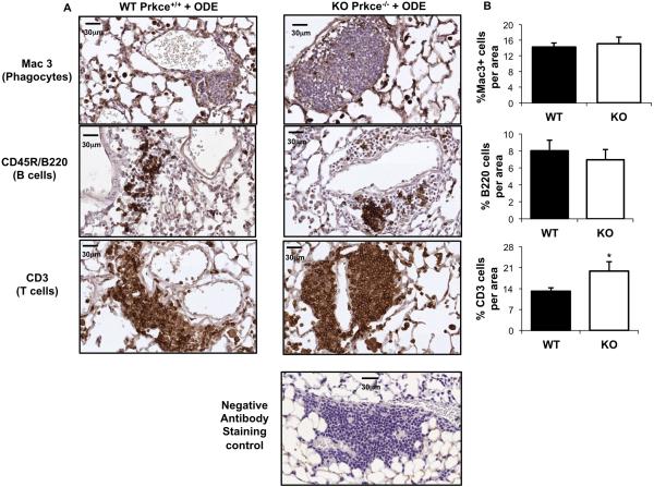
Figure 4. Repetitive organic dust exposure induces the influx of phagocytes, T and B cells in PKCε deficient mice.
Wild-type (WT, Prkce+/+) and PKCε knock-out (KO, Prkce−/−) mice were intranasally treated with ODE (12.5%) daily for 3 weeks. Lung sections (4- to 5-μm thick) were stained with anti-Mac-3 antibody for phagocytes, anti-CD3 antibody for T cells, and anti-murine CD45R/B220 antibody for B cells. Representative lung sections shown depict phagocyte influx into alveolar compartment and predominately T and B cell influx within cellular aggregates in both WT and KO animals repetitively treated with ODE at 40X magnification with scale bar line representing 30 μm. Isotype control antibody staining (negative) control is also shown. B, Quantification of positive staining of mouse lung tissue representing mean ± SEM of 20 fields of representative mouse per group. Statistical significance is denoted by asterisks (*p<0.05) between WT and KO-treated groups.

