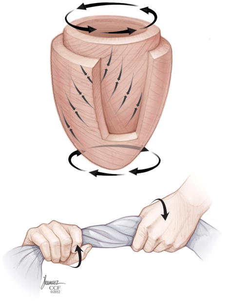
Figure 11.
Model of LV myofiber structure and twist mechanics. The contractile motion of the LV is dictated by the spiral structure of the myocardial fibers. Subendocardial myofibers are wrapped in a right-handed helix, sub-epicardial fibers are wrapped in a left-handed helix, and mid-myocardial fibers are oriented parallel to the circumferential direction. Following a brief clockwise rotation of the apex and opposite (counterclockwise) rotation of the base during isovolumic contraction, the subendocardial and subepicardial fibers shorten concurrently during ejection, causing a “wringing” motion of the apex and base in counterclockwise and clockwise directions, respectively, when viewed from the apex. “Reprinted with permission, Cleveland Clinic Center for Medical Art & Photography © 2013. All Rights Reserved.”

