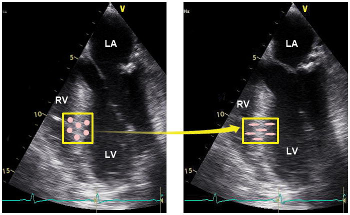
Figure 8.

Myocardial deformation measured by speckle-tracking echocardiography tracks myocardial movement and deformation using the speckles in echocardiographic images. These sequential echocardiographic frames provide an example of the tracking of a unique pattern or “fingerprint” in the myocardial region of interest (yellow box) from frame-to-frame to measure myocardial deformation. The pink circles within the box represent myocardial speckles, which experience shortening in the longitudinal direction and thickening in the transverse direction. RV =right ventricle, LV =left ventricle, LA =left atrium. “Reprinted with permission, Cleveland Clinic Center for Medical Art & Photography © 2013. All Rights Reserved.”
