Fig. 5.
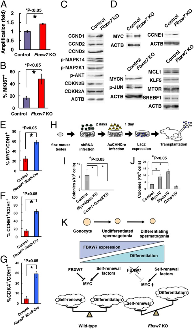
Characterization of Fbxw7 KO GS cells for target identification. (A) Enhanced proliferation of GS cells after Cre transfection. After overnight incubation with AxCANCre, virus supernatant was removed, and cells were replated in a new dish. Cell number was determined 3 d after replating. AxCANLacZ was used as a control (n = 4). Results of two experiments. (B) Quantification of GS cells expressing MKI67. At least 732 cells in four different fields were counted. (C) Western blot analysis of molecules involved in proliferation of GS cells. (D) Western blot analysis of FBXW7 substrates. (E) Expression of MYC in CDH1+ cells in Fbxw7f/f Stra8-Cre testes. Fifteen tubules were counted. (F) Expression of CCNE1 in CDH1+ cells in Fbxw7f/f Stra8-Cre testes. Fifteen tubules were counted. (G) Expression of CDK4 in CDH1 cells in Fbxw7f/f Stra8-Cre testes. At least 8 tubules were counted. (H) Experimental procedure. Immature testis cells from Fbxw7f/f mice were infected with lentiviruses expressing shRNA against Myc/Mycn or Ccne1/Ccne2. Culture medium was changed on the next day after infection. Two days after shRNA infection, cells were infected with AxCANCre and transplanted 24 h after AxCANCre infection. (I) Colony counts after depleting the indicated genes (n = 8). Results of two experiments. (J) Colony counts after overexpression of indicated genes and transplantation of green mouse testis cells. Results of two experiments (n = 12). (K) Summary of experiments. Loss of FBXW7 increases self-renewal by up-regulation of MYC and suppresses differentiation.

