Fig. 5.
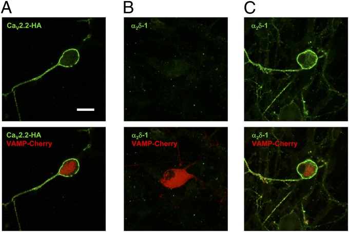
Cell-surface localization of CaV2.2 and α2δ-1 in DRG neurons. Cell-surface expression of CaV2.2-HA (A) and α2δ-1 (B and C) in nonpermeabilized DRG neurons transfected with CaV2.2-HA/α2δ-1/β1b and VAMP-mCherry. Transfected cells were identified by VAMP-mCherry (red). (Lower) Merged images). (A) CaV2.2-HA immunostaining (green); 58/71 mCherry-positive DRG examined (81.7%) had surface HA signal in this condition. (B) α2δ-1 immunostaining (green); 0/20 mCherry-positive DRG had surface α2δ-1 signal. (C) α2δ-1 immunostaining (green) after antigen retrieval; 52/62 mCherry-positive DRG (85.5%) had surface α2δ-1 signal in this condition. (Scale bar, 20 μm.) Representative of two separate transfections.

