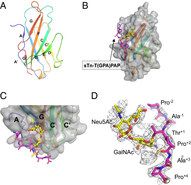
Fig. 2.
Structures of PILRα and its sTn peptide complex. (A) Cartoon representation of the overall structure of the free human PILRα protein. The secondary structural elements are rainbow colored with the N terminus in blue and the C terminus in red. (B) Surface representation of the PILRα-sTn peptide complex. The sialylated O-linked sugar and surrounding peptide are shown as sticks and are colored yellow and magenta, respectively. The same presentation is used in C and D. (C) Close-up view of the sTn-binding region. PILRα is shown as a cartoon model with the surface presentation and coloring as in A. (D) Composite omit map around the sTn peptide (mesh in 1.2σ cutoff).

