
Fig. 3.
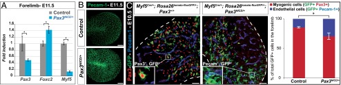
Increase of the number of endothelial cells derived from the somite, in Pax3NICD/+ embryos. (A) qRT-PCR analysis of transcripts of Pax3, Foxc2, and Myf5 compared with gapdh, on forelimbs of Pax3NICD/+ embryos, at E11.5, compared with the controls (Pax3+/+), taken as 1. *P < 0.05, error bars indicate SEM (n = 3 embryos). (B) Whole-mount immunostaining with an antibody to Pecam-1 on the forelimbs of control (Pax3IRES-nLacZ/+) and Pax3NICD/+ embryos, at E11.5 showing the 3D projection (mean value) of the images obtained with confocal sectioning of the limb buds (Scale bar, 100 μm). (C) Immunohistochemistry on sections from control (Pax3+/+;Myf5Cre/+;Rosa26tomato-floxGFP/+) and Pax3NICD/+;Myf5Cre/+;Rosa26tomato-floxGFP/+ embryos, with Pax3 (red), Pecam-1 (blue), and GFP (green) antibodies, at E10.5, at the forelimb level. Insets show enlargements of GFP+Pax3+ (Left) and GFP+Pecam-1+ (Right) cells (Scale bar, 50 μm). The graphs represent quantification of the contribution of somitic cells (GFP+) in the myogenic lineage (GFP+ cells labeled by Pax3) and in the endothelial lineage (GFP+ cells labeled by Pecam-1), expressed as a percentage of total GFP+ cells. *P < 0.05, error bars indicate SEM (n = 3 embryos).