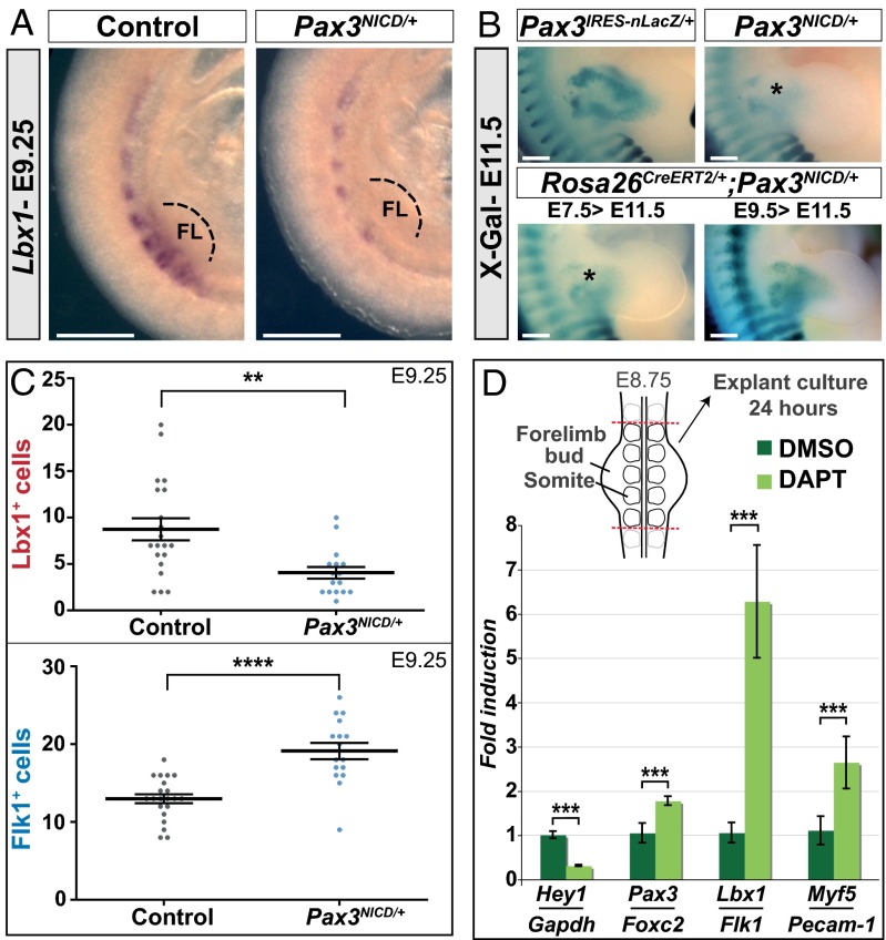
Fig. 4.
Endothelial, versus myogenic cell fate in the somite is promoted by the Notch pathway. (A) Whole-mount in situ hybridization for Lbx1 transcripts on control (Pax3+/+) and Pax3NICD/+ embryos at E9.25. Close ups at the forelimb (FL) level are shown (Scale bar, 500 μm). (B) X-Gal staining of Pax3IRES-nLacZ/+, Pax3NICD/+ and Pax3NICD/+;Rosa26CreERT2/+ embryos, at E11.5. Enlargements at forelimb level are shown. Tamoxifen was injected into pregnant mice at E7.5 or E9.5, as indicated (Scale bar, 2 mm). (C) Quantification of the total number of endothelial progenitors (Flk1+) and myogenic progenitors (Lbx1+) in the hypaxial region at E9.25. **P < 0.01 and ****P < 0.0001. Error bars indicate SEM (n > 16 limb bud sections). (D, Upper) A schema of the explant procedure. (Lower) qRT-PCR results expressed as ratios of Hey1/Gapdh, Pax3/Foxc2, Lbx1/Flk1, and Myf5/Pecam-1 transcripts on explants after 24 h of culture, in DMSO and DAPT (50 µM). ***P < 0.001, error bars indicate SEM (pool of n > 2 explants by experiment, n > 3 experiments).

