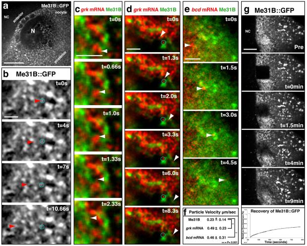
Figure 2. Dynamics of grk, bcd and Me31B particles in live oocytes.
(a) Low magnification wide-field image of the DA corner of a stage 8-9 egg chamber expressing Me31B::GFP. (b) Me31B::GFP expressing oocytes show small faint dynamic particles of Me31B (red arrowheads) moving between and fusing together with other often larger, bright and static (dashed cyan circles) Me31B bodies. (Supplemental Movie 1, 2). (c,d) grk*mCherry and Me31B::GFP expressing oocytes show grk mRNA particles (white arrowheads) moving independently of Me31B (Supplemental Movie 3). (d) Dynamic particles of grk (white arrowhead) are visualized docking and remaining on the edge of Me31B rich zones. Other small grk particles are seen in association with the Me31B throughout the time course (dashed cyan circles) (Supplemental Movie 4). (e) bcd*RFP and Me31B::GFP expressing oocytes show bcd particles (white arrowheads) moving independently of Me31B. (Supplemental Movie 5). (f) Average particle velocities for dynamic Me31B (n=30), grk (n=37) and bcd (n=31) particles in μm/second +/− SEM. P-values from student’s t-tests (two tails), P<0.001. (g) Fluorescence Recovery After Photobleaching of Me31B::GFP at the DA corner shows recovery to approximately 50% of total fluorescence with a half time of 4 minutes (n= 5). NC, Nurse Cell; N, nucleus. Scale bars, 10μm (a); 2μm (b-e); 20μm (g).

