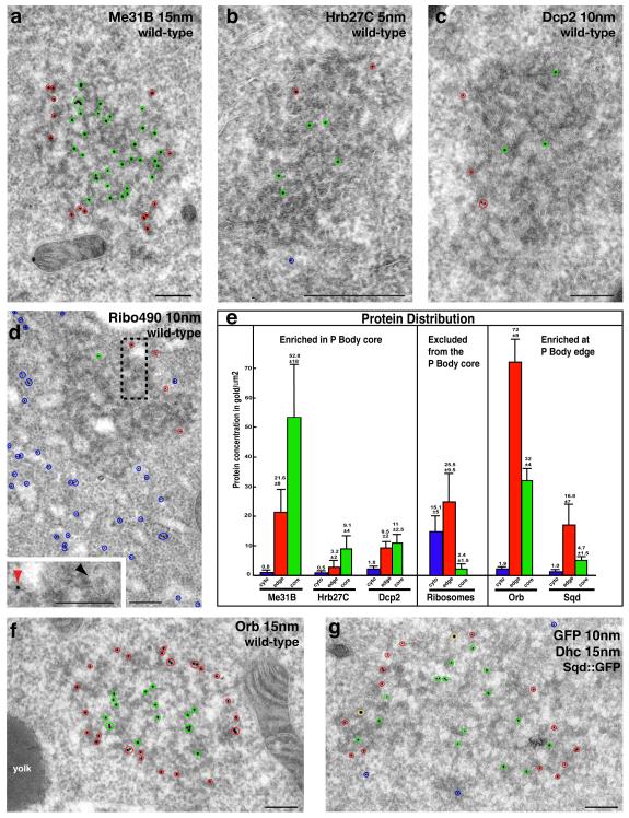
Figure 3. Oocyte P bodies exhibit zones differentially enriched in RNA associated proteins.
(a-d,f,g) IEM localization of RNA associated proteins in stage 9 oocytes at the DA corner. Protein in the core of the electron dense P bodies indicated by green circles, at the edge of P bodies by red circles and in the cytoplasm by blue circles. (a) WT egg chamber, anti-Me31B (15nm) enriched inside P bodies. (b) Hrb27C (5nm) predominantly inside of P bodies. (c) Dcp2 (10nm) inside and at the edge of P bodies. (d) WT egg chamber, anti-Ribo 490 (10nm) shows ribosomes predominantly in the cytoplasm some at the edge but mostly excluded from inside of P bodies. The pool of cytoplasmic ribosomes corresponds to polysomes and those present on the ER membrane (Rough ER), some of which is present near the edge of a P body (black dashed box, inset: red arrowhead). Smooth ER is detected inside of the P body (inset: black arrowhead). (e) Graph showing protein concentration in gold/μm2 in the cytoplasm, edge of P bodies and inside of P bodies; proteins are organized into three distinct categories. Error bars are ± standard deviation (between n=10-15 scans in each case). (f) WT egg chamber, anti-orb (15nm) enriched at the edge of P bodies. (g) Sqd-GFP in Sqd::GFP expressing egg chamber using anti-GFP (10nm) and anti-Dhc (15nm, yellow circles). Sqd is enriched inside compared to at the edge of P bodies. Scale bars, 200nm (a-d,f,g).

