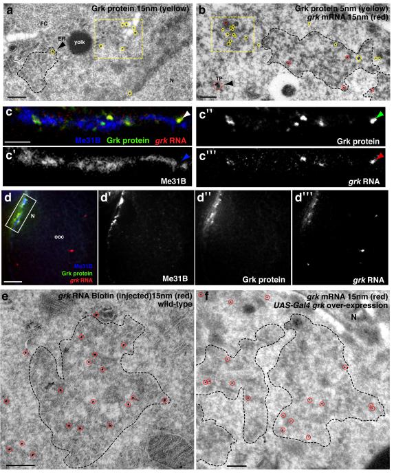
Figure 4. RNA translation fate is regulated through association with P bodies.
(a) IEM using anti-Grk (15nm, yellow circles) on a WT stage 9 egg chamber shows Grk protein highly enriched in the tER-Golgi unit (yellow dashed box) and detected on ER (black arrowhead) at the edge of a P body (black dashed line). (b) ISH-IEM detection of grk mRNA and Grk protein on a WT stage 9 egg chamber shows grk mRNA (15nm, red circles) mostly present at the edge of the P body (black dashed line) and Grk protein (5nm, yellow circles) on a tER-Golgi unit (yellow dashed box) but also present at the edge of the P body (black dashed line). Note the grk mRNA transport particle (dashed red circle) does not contain any detectable Grk protein (black arrowhead). (c) Injection of in vitro synthesized grk RNA (mixed solution of approximately one part Alexa dye labeled and 4 parts unlabeled) into a grk null egg chamber expressing Me31B::GFP. Images shown are a 4μm projection. Localization and translation was allowed for 40 minutes before the egg chamber was fixed and labeled with anti-Grk. (c,c″) Grk protein (green) and (c,c‴) grk RNA (red) colocalize, but do not colocalize with (c,c′) Me31B (blue). Instead, grk RNA and protein interdigitates with Me31B (arrowheads). (d-d‴) Low magnification of panel c. Injected grk RNA is localized to the dorsoanterior corner where it is translated. (e) IEM on a WT stage 8/9 egg chamber injected with a high concentration of grk RNA Biotin (500 ng/μl). Injected RNA (15nm, red circles) is detected at the core of and at the edge of the P body (dashed black line). (f) IEM on a stage 8/9 egg chamber over-expressing grk mRNA using the UAS-Gal4 system. grk mRNA (15nm) is enriched at the core of and at the edge of the P body (dashed black line). FC, follicle cell; TP, transport particle; ER, endoplasmic reticulum; N, nucleus. Scale bars, 200nm (a,b,e,f); 3μm (c); 10μm (d).

