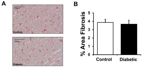
Figure 5. Quantification of interstitial fibrosis in LV rabbit epicardial tissue.

(A.) Representative examples of stained sections (red = collagen). (B.) Quantification of % area fibrosis in control (N=7) and diabetic (N=7) rabbit samples.

(A.) Representative examples of stained sections (red = collagen). (B.) Quantification of % area fibrosis in control (N=7) and diabetic (N=7) rabbit samples.