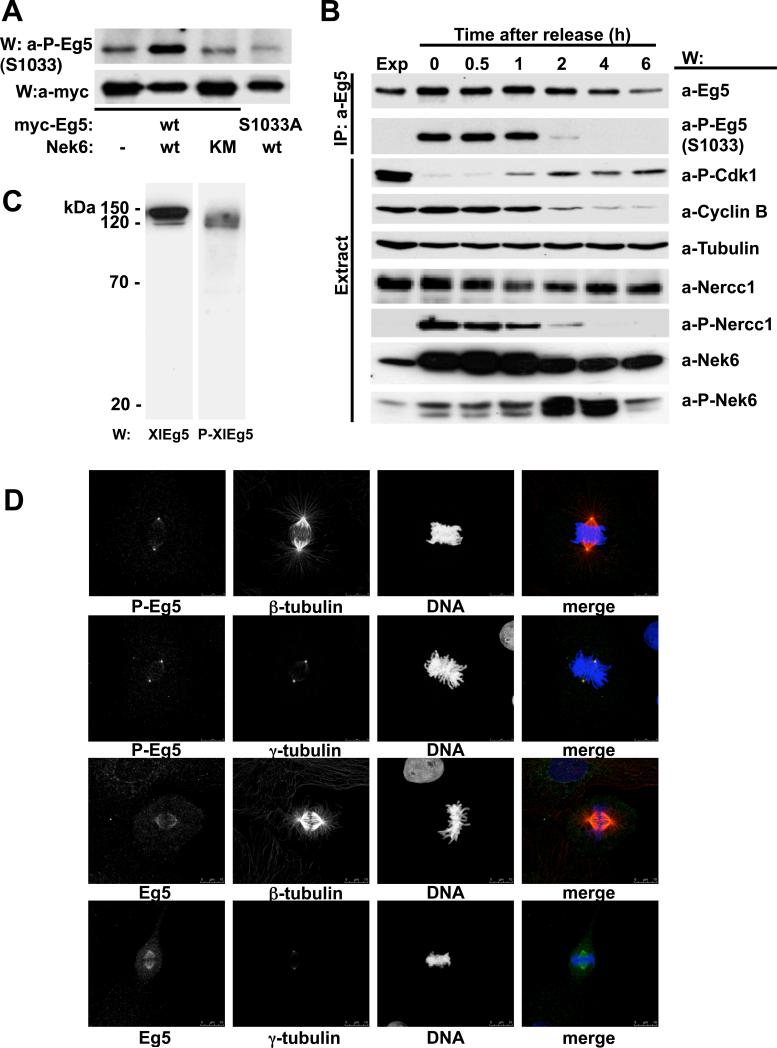
Figure 3. Eg5[Ser1033] phosphorylation in vivo.
(A) U2OS cells were cotransfected with either myc-Eg5 wt (lanes 1-3) or myc-Eg5[S1033A] (lane 4) and empty plasmid (lane1), FLAG-Nek6 wt (lane 2 and 4) or FLAG-Nek6 [K74M,K75M] (lane 3). 24 hours later, anti-Myc-immunoprecipitates were subjected to immunoblot with anti-Myc (lower panel) and anti-Eg5[Ser1033-P] antibodies (top panel). (B) U2OS cells growing exponentially were either untreated (first lane, Exp) or arrested with nocodazole (0.25mM overnight; lane 2); arrested cells were allowed to exit mitosis in nocodazole-free medium and extracted at the times indicated (lanes 3 to 7). Eg5 immunoprecipitates (top two rows) and cell extracts (bottom seven rows) were analyzed by immunoblot with the antibodies indicated (a-P-Eg5(S1033), anti-Eg5[serine1033P]; a-P-Cdk1, anti-Cdk1[Tyr15P]; a-P-Nercc1, anti-Nercc1[Thr210P]; a-P-Nek6, anti-Nek6[Ser206P]. (C) Extracts from XL177 Xenopus laevis cells were resolved by SDS-PAGE and immunoblotted with either anti-XlEg5 (left) or anti-XlEg5[Ser1046-P] (right). (D) XL177 cells growing exponentially were fixed and stained with antibodies to XlEg5 (lower two rows), XlEg5[Ser1046P] (upper two rows), and either β- or γ-tubulin as indicated. DNA is stained with DAPI. Representative cells in metaphase are shown; bar, 10 μm.

