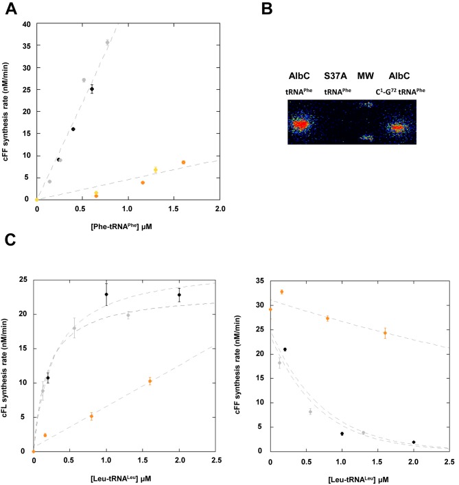
Figure 3.
(A) cFF-synthesizing activity of AlbC using either Phe-tRNAPhe purified from E. coli (black/grey) or Phe C1-G72 tRNAPhe (orange/yellow). The points reported are the result of two independent experiments. Error bars show the uncertainty on measurement. Curves are drawn for clarity and are not representative of kinetic models. Enzymatic measurements were performed as described in ‘Materials and Methods’ with 50 nM AlbC. (B) Covalent labelling of AlbC and S37A by [14C]Phe-tRNAPhe or [14C]Phe C1-G72 tRNAPhe. Enzymes were incubated with labelled aa-tRNA, as described in ‘Materials and Methods’, separated on SDS–PAGE, then transferred onto a PVDF membrane that was analysed with a radioimager. (C) Rate of formation of cFL (left panel) and cFF (right panel) by AlbC using tRNALeuCAG obtained by in vitro transcription (black), tRNALeuCAG purified from E. coli (grey) or Leu C1-G72 tRNALeuCAG (orange). Kinetics of synthesis were determined as described in Figure 1. Curves are drawn for clarity and are not representative of kinetic models.

