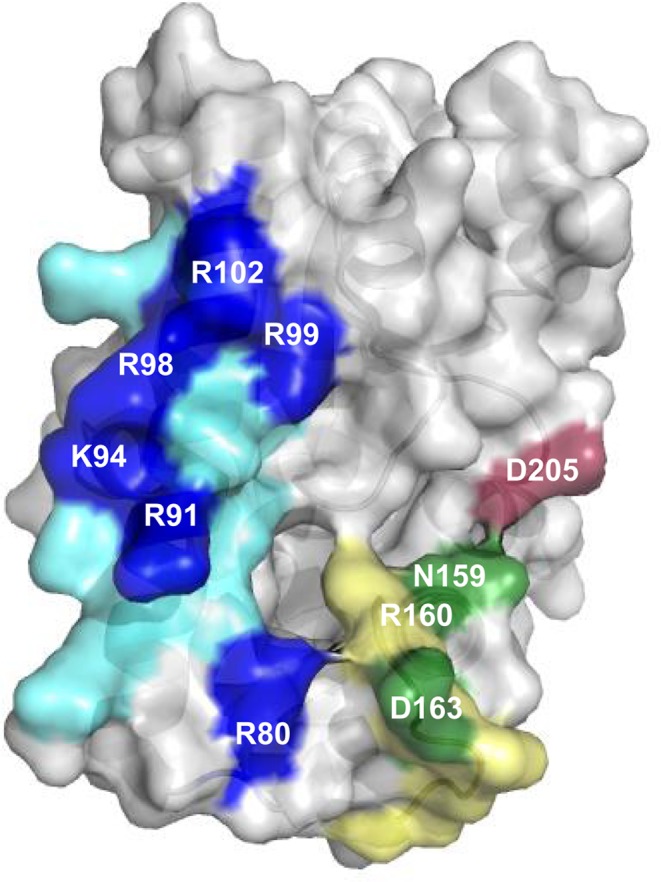
Figure 5.

AlbC regions involved in interaction with tRNA substrates. The overall structure of AlbC (PDB, 3OQV) is shown in surface mode. The patch of basic residues located on helix α4 is coloured in blue; the residues substituted in this study are in dark blue. The loop α6–α7 is coloured in green; the residues substituted in this study are in dark green. The residue D205 belonging to the loop β6- α8 is coloured in dark red.
