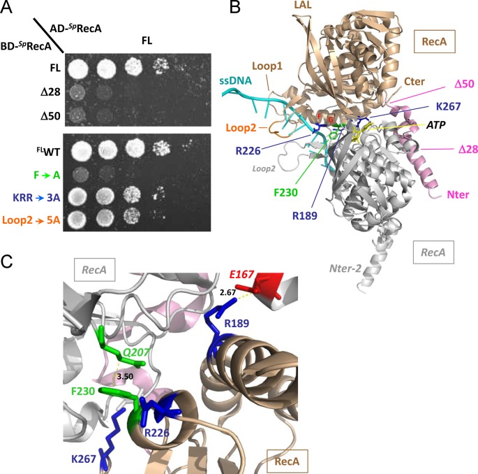
Figure 3.
RecA−RecA interaction. (A) Yeast strains expressing wild-type SpRecA fused to Gal4 activation domain (AD-SpRecA) and SpRecA variants as Gal4 binding domain fusions (BD-SpRecA) were spotted as a series of 1/5th dilutions on selective medium lacking histidine. Plates were incubated for 5 days at 28°C. (B) Ribbon schematic representation of the SpRecA−SpRecA dimer modeled from the X-ray structure of EcRecA−ssDNA nucleo-filament (PDB ID: 3CMW (2)). The two monomers are in gold and gray, the ssDNA is in cyan. The color code for the residues concerned in the DprA interaction is the same as in Figure 2. (C) The RecA−RecA interaction zone is enlarged to show the residues (represented using the same color code as in (B)) involved in the interaction. Hydrogen bonds and salt bridges are represented by dashed lines, and distances between residues are indicated in Å. RecAF230 is in apolar contact with RecAQ207 of the other subunit.

