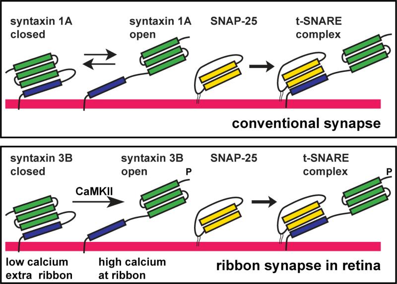
Figure 10. Model of the regulation of syntaxin 3B in ribbon synapses of the retina.
In conventional synapses syntaxin 1A exists in two conformations: “open” and “closed”. The “closed” conformation has the Habc domain (green) folding back on the SNARE domain (blue) and cannot bind to SNAP-25 to form a t-SNARE complex. We propose that syntaxin 3B in ribbon synapses of the retina exists mainly in the closed conformation. Activation of CaMKII by elevated calcium leads to the phosporylation of syntaxin 3B in the proximity of the ribbon. The phosphorylation of syntaxin 3B stabilizes the open conformation that can then efficiently form a t-SNARE complex and increase synaptic vesicle exocytosis.

