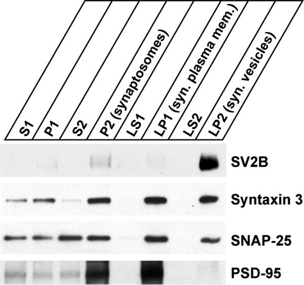
Figure 5. Syntaxin 3B is associated with the synaptic plasma membrane.
Subcellular fractionation of mouse retina was performed to analyze the distribution of syntaxin 3B. Synaptosomes (P2) were isolated from mouse retina by differential centrifugation. The synaptosomes were then hypo-osmotically lysed and separated into synaptic plasma membrane fraction (LP1), synaptic vesicle fraction (LP2) and soluble presynaptic protein fractions (LS1 and LS2). Equal protein amounts were analyzed by SDS-PAGE and western blotting using specific antibodies directed against SV2B (ribbon synaptic vesicle marker), syntaxin 3, SNAP-25 and PSD-95 (postsynaptic density protein).

