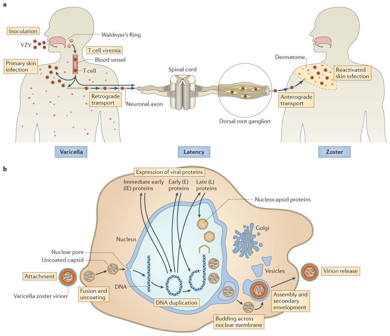
Figure 1. VZV life cycle and replication.
a | Model of the varicella zoster virus (VZV) life cycle. VZV infects the human host when virus particles reach mucosal epithelial sites of entry. Local replication is followed by spread to tonsils and other regional lymphoid tissues, where VZV gains access to T cells. Infected T cells then deliver the virus to cutaneous sites of replication. VZV establishes latency in sensory ganglia after transport to neuronal nuclei along neuronal axons or by viraemia. Reactivation from latency enables a second phase of replication to occur in skin, which typically causes lesions in the dermatome that is innervated by the affected sensory ganglion. b | Model of VZV replication. Enveloped VZV particles attach to cell membranes, fuse and release tegument proteins. Uncoated capsids dock at nuclear pores, where genomic DNA is injected into the nucleus and circularizes. On the basis of events that have been documented in herpes simplex virus 1 (HSV-1) replication, immediate-early genes are expressed, followed by early and late genes. Nucleocapsids are assembled and package newly synthesized genomic DNA, move to the inner nuclear membrane and bud across the nuclear membrane. Capsids enter the cytoplasm, and virion glycoproteins mature in the trans-Golgi region and tegument proteins assemble in vesicles; capsids undergo secondary envelopment and are transported to cell surfaces, where newly assembled virus particles are released.

