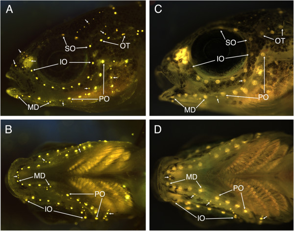
Figure 6.

Fluorescent imaging of neuromasts in Tramitichromis sp. and Aulonocara stuartgranti using 4-di-2-ASP. A, B) Lateral and ventral views of Tramitichromis sp. (11 mm SL), with arrows pointing to first and last canal neuromasts in the supraorbital (SO), infraorbital (IO), preopercular (PO), mandibular (MD) and otic/post-otic (OT) series. Small arrows point to various groups of superficial neuromasts (see [58] for naming). C, D) Lateral and ventral views of Aulonocara stuartgranti (11.5 mm SL) labeled as in A and B. In this individual (the same as visualized with SEM in Figure 7D), the fluorescent label illuminates the entire neuromast (hair cells and surrounding support cells) revealing their diamond shape (as in Figure 7L).
