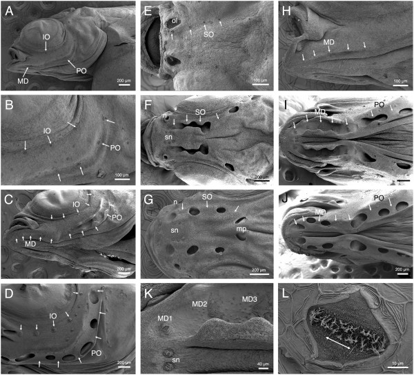
Figure 7.

Neuromasts and canal morphogenesis in Aulonocara stuartgranti and Tramitichromis sp. visualized with SEM. All SEM images are of late stage larvae and juveniles of Aulonocara, with the exception of C, which is of Tramitichromis. Arrows indicate presumptive CNs or position of CNs after canal enclosure; rostral to left in all images; see Figure 2 and 8 for definitions of St. 1 to St. 4. A) Infraorbital (IO), mandibular (MD) and preopercular (PO) presumptive CNs (canal neuromasts) in yolk sac larva (y, yolk; 7.5 mm SL). B) Close-up of A showing IO CNs at St. I, PO CNs at St. IIa. C)Tramitichromis sp. (7.5 mm SL): IO CNs at St. I, MD (MD1 to MD5) at St. I, and PO CNs at St. I or St. IIb. D) IO CNs at St. I and PO CNs at St. III/IV (11.5 mm; same individual as in Figure 6C,D). Other neuromasts are small SNs (superficial neuromasts) that remain on skin. E) Four supraorbital (SO) CNs; SO1 is medial to olfactory organ (ol) (6.0 mm SL). F) SO canal with SO1 at St. I, medial to naris (n), SO2 and SO3 at St. IIb; SO 4 and SO5 at St. III/IV; SNs visible between SO canals (sn; 9.5 mm SL). G) SO canal at St. III/IV, arrows indicate SO1 to SO3; two pores caudal to position of right and left SO3 CNs will fuse to form medial pore (mp) (10 mm SL). H) Mandibular CNs (MD1 to MD5) at St. IIa (7 mm SL). I) MD canal with MD1 at St. IIa, MD2 to 4 at St. IIb, and MD5 at St. IIa. First two PO CNs are at St. IIb and St. III/IV (11 mm SL). J) MD and PO canals enclosed (St. III/IV), arrows show MD1 to MD5 and first two PO CNs (12 mm SL). K) Close-up of left MD canal in I, showing MD1 at St. IIa, and MD2 and MD3 at St. IIb; small SNs (sn) are round in contrast to diamond-shaped canal neuromasts (11 mm SL). L) Close-up of diamond-shaped CN, MD1, showing location of sensory hair cells in sensory strip elongated parallel to physiological orientation of hair cells and canal axis (double-headed arrow; 10 mm SL).
