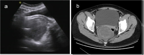
Figure 1.

Image feature of tumor pre-operation. (a) Transvaginal ultrasonography shows an oval, moderately echoic mass in close proximity to the cul-de-sac of Douglas. (b) Pelvic CT scan shows a well-circumscribed tumor of homogeneous intensity located anteroinferior to the sacrum.
