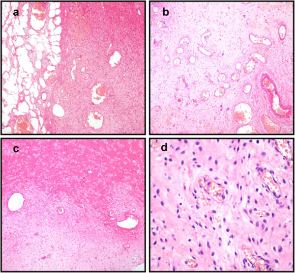
Figure 2.

Histopathological examination of tumor post-operation. (a) The tumor is well demarcated from the surrounding fat tissues. (b) Abundant thin-walled blood vessels can be seen in the tumor. (c) The stroma of the tumor is hyalinized or edematous, and appears hypocellular in some areas. (d) The tumor is composed of bland, plump, spindle-shaped or oval cells that are frequently aggregated around thin-walled blood vessels (H&E: 100 x).
