Figure 5.

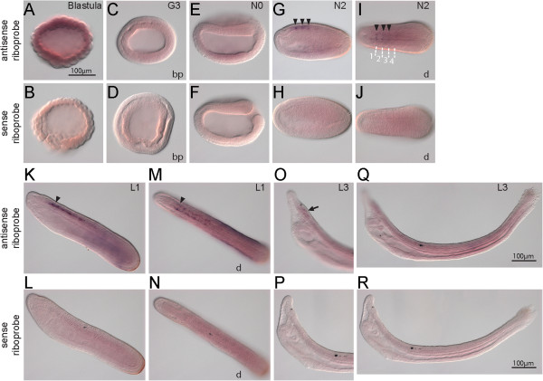
Expression of BfNcoa.In situ hybridization of BfNcoa with antisense probe and with sense probe. (A-F) Ubiquitous expression is shown from blastula to early neurula. (G-N) From mid-neurula, tissue-specific signal is detected in some paired cell groups in the anterior central nervous system (arrowheads). (O,P) At the larval stage, stronger expression is observed in the cerebral vesicle (arrow), and weaker expression is observed in the neural tube. (Q,R) No apparent expression is found in the gut. The scale bar in panel A applies to panels A-P. Blastoporal views (bp) and dorsal views (d) are labeled, and unlabeled panels are lateral views. In E-J, anterior is to the left; in K-R, anterior is to the upper left. Boundaries of somites are depicted in I.
