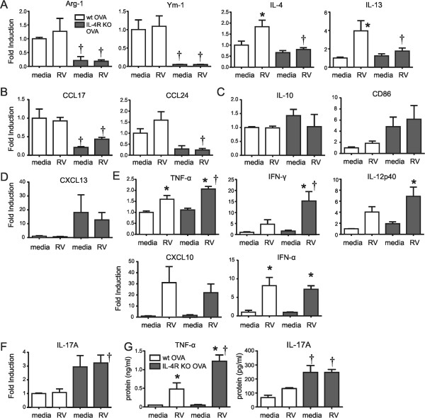
Figure 4.

Differential cytokine expression in RV-stimulated macrophages from OVA-treated wild-type and IL-4R KO mice. Macrophages were collected from the BAL of OVA-treated wild-type or IL-4RKO mice. Macrophages were selected by allowing adherence to plastic for 2 h. Macrophages were treated with medium or RV (multiplicity of infection, 5) for 1.5 hours. Cells were collected 8 h or 24 h after infection for RNA and protein analysis. Gene expression in macrophage was measured by qPCR. Shown are M2 surface markers and the type 2 cytokines IL-4 and IL-13 (A), the M2a markers CCL17 and CCL24 (B), the M2b markers IL-10 and CD68 (C), the M2c marker CXCL13 (D), the classical M1 markers TNF-α, IFN-γ, IL-12p40 and CXCL10, IFN-α (E) and IL-17A (F). (G) TNF-α and IL-17A protein levels were assessed with ELISA. (Data represent three independent experiments, mean ± SEM, n = 3-8 each group, *different from sham, P < 0.05, one-way ANOVA; †different from wild type, P < 0.05, one-way ANOVA).
