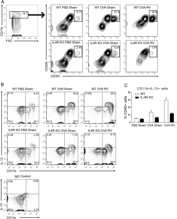
Figure 5.

Differential expansion of CD206+ CD301+ M2-polarized macrophages and IL-13 production in wild-type and IL-4R KO mice. Eight-week old wild-type or IL-4R KO mice were treated with PBS or OVA by intraperitoneal injection (days 0, 7) and intranasal installation (days 12, 13). Mice were intranasally inoculated with sham or RV on day 14. Lungs were harvested and minced in collagenase IV solution. (A) Cells were stained with antibodies against macrophage surface markers and assessed with flow cytometric analysis. CD206- and CD301-double positive cells in the CD11b+ cell fraction are shown. (B) Cells were incubated with cell stimulation cocktail for 5 h, stained, and analyzed with flow cytometric method. Expression of CD11b and IL-13 was analyzed among CD45+ cells. The numbers represent the percentage of cells within each quadrant. For the control, IgG was substituted for all antibodies. (C) The percentage of CD11b + IL-13+ cells were shown among CD45+ fraction. N = 4, *different from PBS sham group, p < 0.05, ANOVA.
