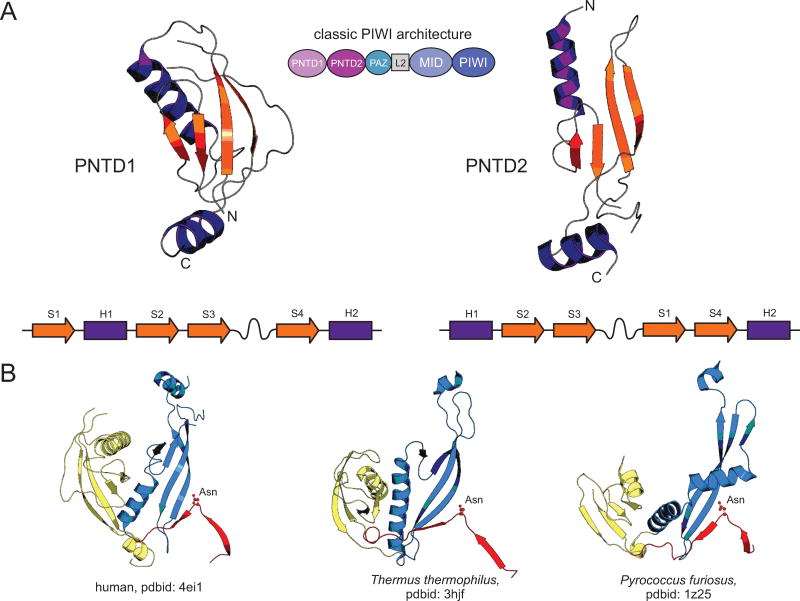
Figure 4.
Structural anatomy of PIWI N-terminal domain 1 (PNTD1) and PIWI N-terminal domain 2 (PNTD2). (A) Cartoon rendering and topological diagrams of the PNTD1 and PNTD2 domains. PNTD2 has previously been referred to as the Linker-1 domain, however, as these renderings make clear, the PNTD2 domain emerged via duplication and circular permutation from the PNTD1 domain, forming a single N-terminal module which is evolutionary present across all three superkingdoms of Life and is found in architectures outside of the well-studied classical PIWI architecture depicted in the center of the figure (see also Fig. 5A). β-strands are colored in orange, α-helices are colored in purple, and extended loop regions are colored in grey. N- and C-termini are labeled with “N” and “C”, respectively. (B) Cartoon renderings of the PNTD1/PNTD2 dyad along with the N-terminal leader region containing the well-conserved asparagine residue. Three protein representatives from the three superkingdoms of Life are depicted, labeled by species and pdbid at the bottom. PNTD1 is colored in yellow, PNTD2 is colored in blue, and the leader region is colored in red. The asparagine residue is rendered as a ball and stick, colored in red, and labeled as “Asn”.

