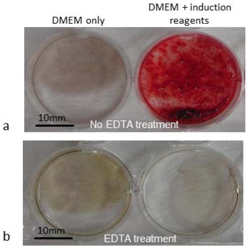
Fig. 1.

Osteogenic induction of DFSCs resulted in formation of calcium deposits as determined by Alizarin Red staining. (a) Alizarin Red positive staining was seen only when cells were cultured in the DMEM medium supplemented with induction reagents (right), and no Alizarin Red staining was seen in the DFSCs cultured in DMEM medium without induction reagents (left). (b) 10% EDTA treatment of the cultures prior to Alizarin Red staining completely removed the deposits and resulted in no staining in the induced cells (right) similar to the non-induced cells (left).
