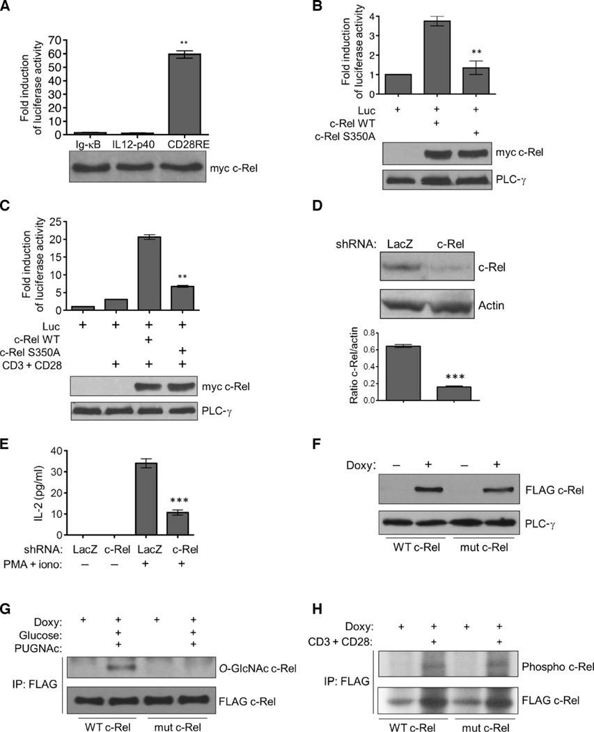
Fig. 3. Mutating the O-GlcNAcylation site blocks the activity, but not the phosphorylation, of c-Rel.
(A) HEK 293T cells were transfected with plasmid encoding myc-tagged WT c-Rel together with the indicated luciferase reporter plasmids. Luciferase activity was assessed with a dual luciferase assay system. **P < 0.01 for the CD28RE reporter compared to the Ig-κB or IL12-p40 reporters. (B and C) Jurkat cells were transfected with plasmids encoding myc-tagged WT c-Rel or the S350Amutant c-Rel together with the CD28RE luciferase reporter plasmid (Luc). (C) Twenty-four hours after transfection, cells were stimulated with anti-CD3 and anti-CD28 antibodies for 4 hours. (B and C) Luciferase activity was assessed as described in (A). **P < 0.01, when comparing the S350A mutant c-Rel to WT c-Rel. (A to C) Data in the bar graphs are means ± SEM of three independent experiments. Bottom: Western blotting analysis of total cell lysates with antibodies against the myc tag or phospholipase C-γ (PLC-γ). Blots are representative of three independent experiments. (D) shRNA-mediated knockdown of endogenous c-Rel in Jurkat T-REx cells. Bottom: Densitometric quantification of c-Rel amounts relative to those of actin. Data are means ± SEM from three independent experiments; the Western blots are from one representative experiment. ***P < 0.001. (E) Jurkat T-REx cells were treated with PMA (50 ng/ml) and ionomycin (250 ng/ml) for 16 hours, and the amounts of IL-2 secreted into the culture medium were determined by enzyme-linked immunosorbent assay (ELISA). Data are means ± SEM from three independent experiments. ***P < 0.001. (F) Jurkat T-REx cell clones were treated with doxycycline (1 µg/ml) for 22 hours, and the production of WT and mutant c-Rel was determined by Western blotting analysis with an anti-FLAG antibody. PLC-γ was used as a loading control. (G) Jurkat T-REx cells (75 × 106) expressing WT and mutant c-Rel were incubated with doxycycline, then treated as described in Fig. 1B, and subjected to immunoprecipitation and Western blotting analysis with antibodies against the indicated proteins. (H) Cellular phosphorylation of WT and S350A mutant c-Rel. FLAG-tagged c-Rel proteins were immunoprecipitated from 75 × 106 Jurkat T-REx cells and analyzed by autoradiography (top) and Western blotting (bottom). Data in (F) to (H) are representative of three independent experiments.

