
Figure 3.
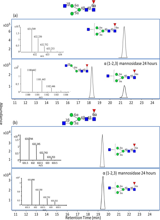
Chromatograms (with MS inset) produced during exoglycosidase sequencing of two isomers with composition N33100 (1261.50 Da, neutral mass). (a) The upper chromatogram shows isomer N33100a before digestion. The representation identifies the uncertainty in the structure. The lower panel includes the ECC of the neutral mass 1261.50 and 1099.45 Da. A single mannose was lost after 24-h digestion with α(1-2,3)mannosidase (MS inset) suggesting an uncapped mannose structure on the 1-3 antenna. The results show that the terminal GlcNAc is on the 1-6 branch. (b) The upper chromatogram shows isomer N3310b before enzyme digestion (MS inset). This compound was analyzed at the same time as the other isomer. There was no loss in signal or new smaller homologue produced when the compound was reacted with α(1-2,3) mannosidase.