
Figure 3.
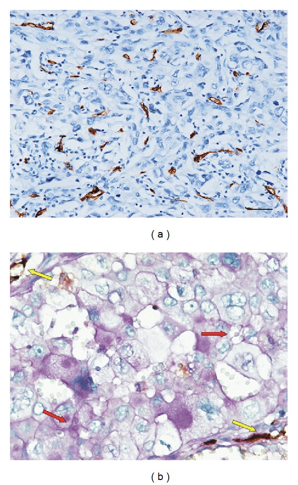
The angiogenesis status in NSCLC. (a) MVD staining for CD34 in NSCLC (immunohistochemical staining, ×200). A hotspot with high MVD was positively stained. (b) CD31/PAS double staining for VM (×400). The VM channel showed a positive expression for PAS but a negative expression for CD31 (red arrow). The endothelial channel showed positive expressions for both CD31 and PAS (yellow arrow).