Fig. 2.
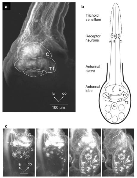
Anterograde labeling of antennal sensory neurons with rhodamine dextran reveals axonal projections to glomeruli in the antennal lobe of Manduca. a Labeling of receptor neurons in long antennal trichoid sensilla shows projections to the three glomeruli of the macroglomerular complex (MGC: C cumulus, T1 toroid-1, T2 toroid-2). b Schematic diagram of receptor neuron projections to the ipsilateral antennal lobe. Projections are shown to all three glomeruli of the macroglomerular complex. Receptor neurons from a given trichoid sensillum project to only one or a combination of two glomeruli in the macroglomerular complex. c Labeling of long trichoid as well as other antennal sensilla shows projections to the MGC and ordinary glomeruli in the AL. Frontal view. Optical sections at different depths from anterior to posterior through the AL from left to right. C cumulus, do dorsal, la lateral, T1 toroid-1, T2 toroid-2. Scale bar 100 μm

