
Figure 2. Pregranulosa cells lose p27 expression and precociously upregulate AMH in Wnt4-null ovaries near birth.
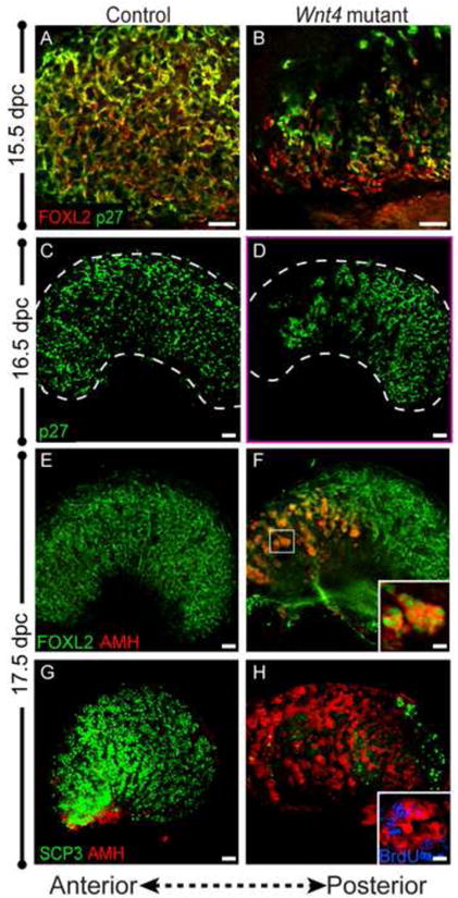
(A,B) At 15.5 dpc, FOXL2 (red) and p27 (green) are co-expressed in pregranulosa cells of control ovaries (A). In Wnt4 mutants, a small group of pregranulosa cells at the anterior of the ovary downregulate p27 (B) (Panels A and B show the anterior end of the ovary). (C,D) By 16.5, the population of pregranulosa cells that downregulated p27 (green) has expanded in an anterior-posterior direction, and continues to do so until birth (data not shown). The border of the ovary is marked by a white dotted line. (E,F) By 17.5 dpc, FOXL2-expressing pregranulosa cells (green) ectopically express AMH (red) in the anterior end of the ovary (see high magnification of boxed region, F, inset). (G,H) AMH-expressing cells (red) are found throughout the ovary near birth, but are excluded from the posterior end of the ovary occupied by meiotic germ cells (SCP3, green). These AMH-positive cells are actively proliferating and are labeled by BrdU (inset in H; blue). Whole mount immunostaining was performed for all samples. Scale bars represent 50 μm in main panels, and 5.5 μm and 2.75 μm in insets in F and H, respectively.