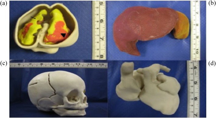
Figure 3.
Rapid prototyping of foetal and neonatal organs from post-mortem MRI data. (a) Foetal brain inside the skull. Cerebrospinal fluid is shown in white with black arrow and intraventricular bleed in grey with black arrowhead. (b) Liver of a newborn. (c) Parietal skull fracture in an infant. (d) Foetal heart.

