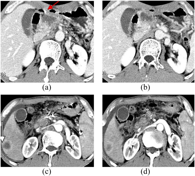
Figure 4.
(a,b) CT of a male patient (62 years old) with upper abdominal pain lasting 1 year. CT examination showed pancreatic mass lesions invading the gastric wall (arrow); the carbohydrate antigen carbohydrate antigen 19-9 was 301 U ml−1; and biopsy verified pancreatic adenocarcinoma. (c,d) On the third day after implantation, upper gastrointestinal bleeding occurred. 2 months later, lesions achieved CR, but hepatic metastasis occurred (see arrows).

