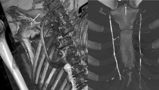
Figure 4.

Normal bone variations useful for comparative identification. (Left image) volume rendering technique reconstruction of skeletal remains. Calcification of the transverse superior ligament of the scapula (arrow). (Right image) Coronal maximum intensity projection reconstruction of the sternum. Presence of a superior episternal bone (arrow).
