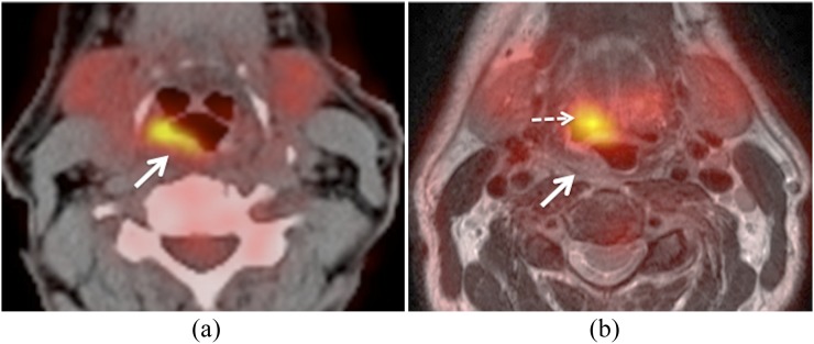
Figure 3.
Positron emission tomography (PET)/MRI and PET/CT obtained for primary staging of squamous cell carcinoma of the hypopharynx. (a) Axial PET/CT image shows a hypermetabolic tumour located in the posterior hypopharyngeal wall (arrow). (b) Corresponding hybrid PET/MRI (fused PET and T2) shows poor data fusion due to patient motion. Note anterior displacement of the PET image in comparison with T2, suggesting hypermetabolic base of the tongue–vallecula tumour (dashed arrow). True location of the tumour in the hypopharynx (arrow).

