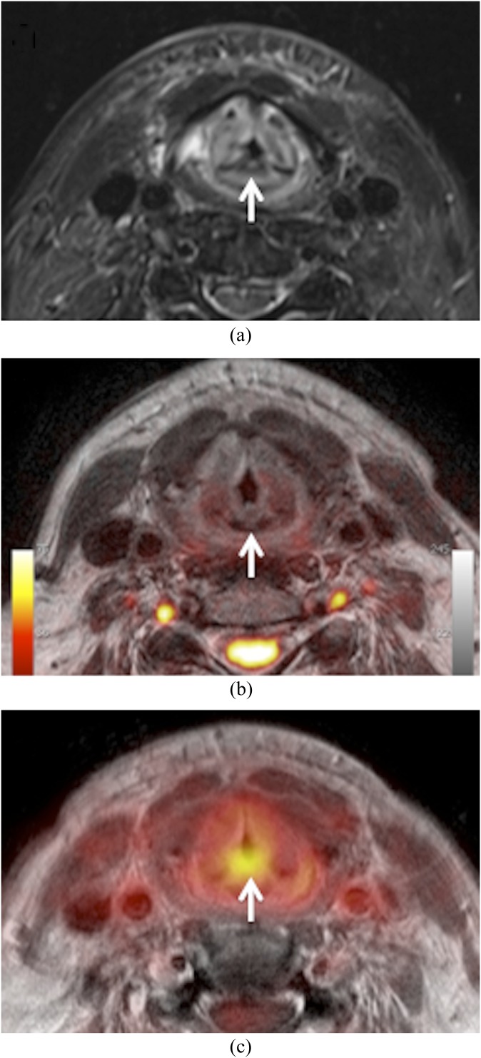
Figure 4.
Hybrid positron emission tomography (PET)/MRI obtained 6 months after radiotherapy of laryngeal squamous cell carcinoma. Clinically, recurrence was suspected. (a) Axial fat-saturated T2 shows diffuse oedema with posterior commissure involvement (arrow). No evidence of recurrence. (b) Fused T2 and b 1000 illustrate absent restriction of diffusivity (arrow). Normal high signal of the spinal cord and nerve roots on b 1000. No major geometric distortion. (c) Fused PET and gadolinium-enhanced T1 show increased 18-fludeoxyglucose uptake (mean standardized uptake value = 3.8; maximum standardized uptake value = 5.2) in the posterior commissure (arrow) suggesting recurrence. Surgical biopsy and follow-up revealed scar tissue.

