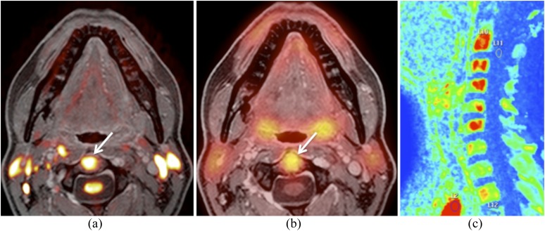
Figure 6.
Images obtained from the same hybrid positron emission tomography (PET)/MRI examination as shown in Figure 5. (a) Fused b 1000 and gadolinium-enhanced water-only Dixon image show restricted diffusivity in the C2 vertebral body (arrow). (b) Fused PET and gadolinium-enhanced water-only Dixon image illustrate increased 18-fludeoxyglucose uptake (mean standardized uptake value = 4.4; maximum standardized uptake value = 6) in the C2 vertebral body (arrow). Similar findings were present in the vertebral bodies of C3–C6 (not shown). The vertebral bodies had been included in the radiation portal. Nevertheless, bone metastases were suspected. (c) Sagittal maximum enhancement perfusion map obtained by dynamic gadolinium-enhanced MRI shows increased vascularization in the vertebral bodies of C2–C6 (in red) supporting the diagnosis of bone metastases. Bone biopsy and follow-up confirmed metastases.

