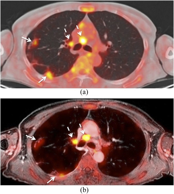
Figure 7.
This patient was a follow-up case of a squamous cell carcinoma of the oropharynx. (a) Axial positron emission tomography (PET)/CT shows metastatic mediastinal lymph nodes (dashed arrows) and metastatic lung nodules (arrows). (b) Corresponding hybrid PET/MRI (fused PET and gadolinium-enhanced water-only Dixon image and slice thickness of 2 mm) shows similar findings. Metastatic mediastinal nodes (dashed arrows). Lung metastases (arrows). Note that lung nodule conspicuity is slightly better on PET/CT than on PET/MRI.

