
Fig. 4.
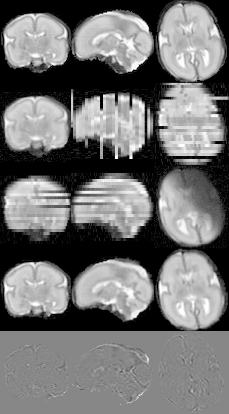
Simulation of fetal brain MRI from a neonatal subject with GA 27 weeks. First row: Neonatal volume. Second row: Simulated coronal stack with 6 slices with large displacement to simulate misregistered outliers. Third row: Simulated transversal stack with three corrupted slices. Fourth row: Reconstruction using six stacks, which include stacks shown in the second and third row, demonstrates the good performance of the method when compared to neonatal volume int the first row. Fifth row: The difference between original and reconstructed image.