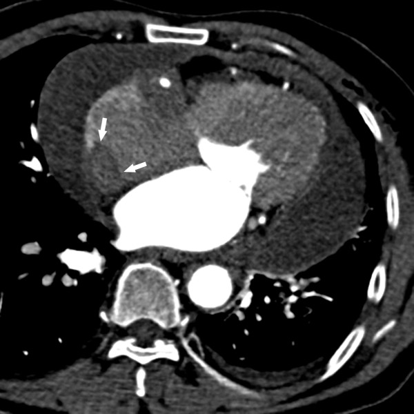
Figure 1.

A contrast-enhanced computed tomography image demonstrating an irregularly shaped, 25.0-mm right atrial mass extending along the right atrial free wall (white arrow); minor left-sided pleural effusion; slight pleural thickening of the right side; and diffuse pericardial effusion.
