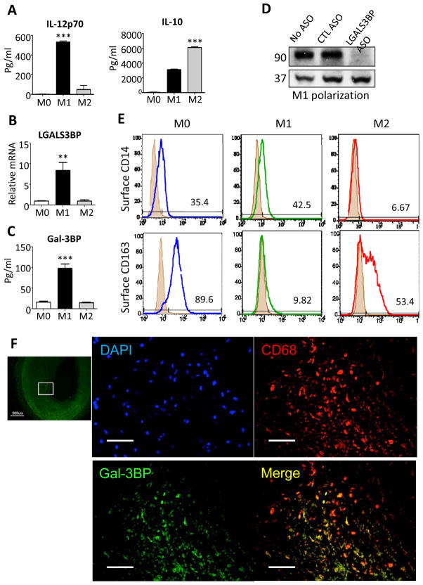
Figure 1. Gal-3BP, CD14 and CD163 expression in human macrophages.
Human blood monocytes (all subsets) were incubated with M-CSF (100 ng/ml) for 6 days to produce monocyte-derived macrophages (M0). These macrophages were incubated with interferon-γ for 1 day to produce M1 or with IL-4 to produce M2 macrophages and then challenged with LPS (10 ng/ml). (A) Level of IL-12p70 and IL-10 in the conditioned media were measured by Elisa (B) Gal-3BP mRNA expression was measured by quantitative RT-PCR, expressed relative to H18S. ** P=0.0056 by paired t test, n=3 donors. (C) Gal-3BP protein production was measured using ELISA (P=0.0008). (D) M1 polarized macrophages were treated with cell-permeable antisense (ASO) against LGALS3BP or control sequence (CTL) for 48hr, conditioned media harvested for immunoblotting with anti-Gal-3BP and anti-ERK2 (loading control) antibodies. (E) CD-14 and CD-163 determined by flow cytometry in M0, M1 and M2 polarized macrophages (representative of 3 healthy blood donors). (F) Human postmortem coronary arteries from patients with coronary artery disease were stained with antibodies against Gal-3BP (FITC, green), CD68 (Texas red), and nuclear stain (DAPI, blue) to assess expression of Gal-3BP in plaque macrophages. Lowest panel shows merge of Gal-3BP and CD68; right panels magnify boxed areas. Top: overview (scale bar = 500 μm), other scale bars = 100 μm

