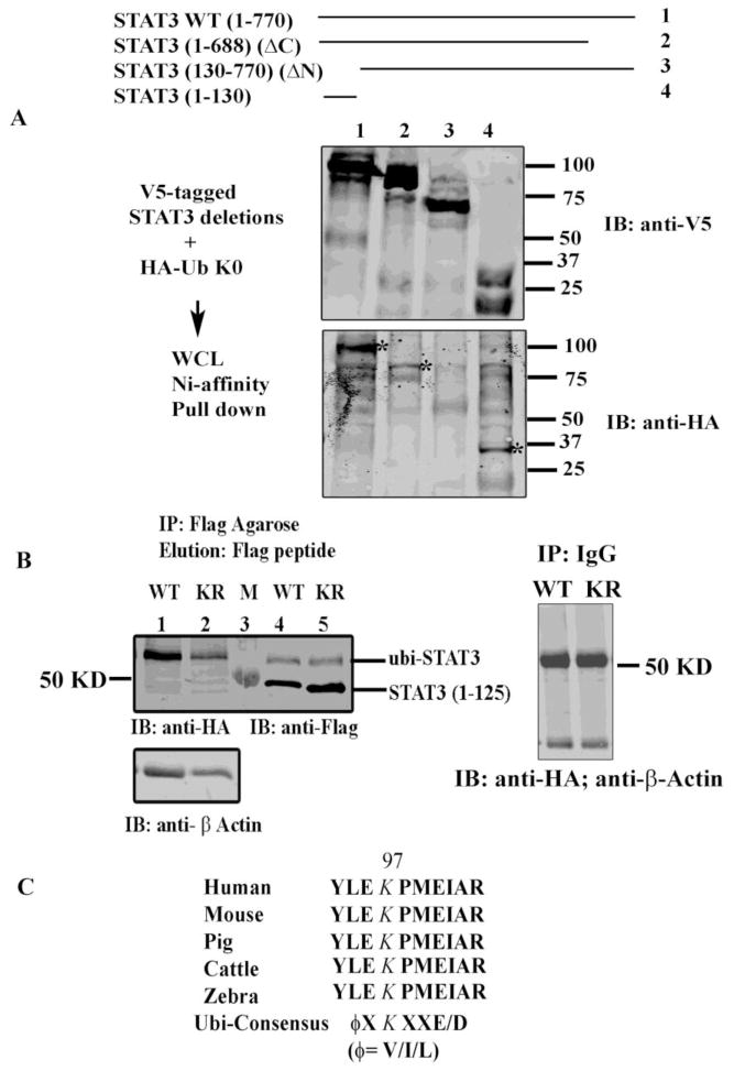
Fig 2.
STAT3 is ubiquitinated on its NH2 terminal domain. (A) HEK293 cells were transfected with the indicated V5-His-tagged STAT3 WT and deletion constructs and HA-ubi-K0. WCL were subjected to Ni-NTA affinity assay and the proteins were resolved and immunoblotted with either anti-V5 Ab (top) or anti-HA Ab (bottom). * represent monomeric ubiquitin conjugated STAT3. (B) HEK 293 cells were transfected with either Flag-tagged STAT3 WT (1-125) or with Flag-tagged K49/87R (1-125) as indicated. WCL were IPed with Flag-agarose beads and proteins were eluted with 3X Flag peptide prior to resolved in SDS-PAGE. Western immunoblot analysis was performed either with anti-HA Ab (left) or with anti-Flag Ab (right). Bottom left panel is the immunoblots of lane 1 and 2 with β-Actin Ab. Right panel shows WCL IP’ed with IgG as a negative control and immunoblotted with anti-HA and β-Actin Ab. M represents Marker lane. (C) ClustalW sequence analysis of STAT3 (1-125) showed conservation of K residue 97 among different mammalian species and contains an ubiquitin/sumoylation consensus sites.

