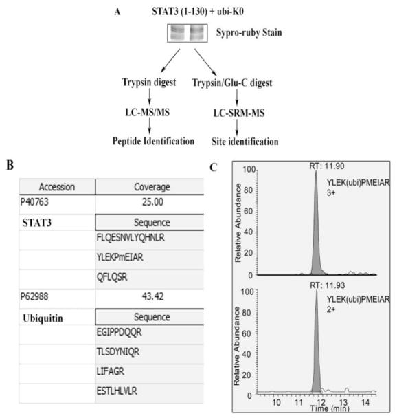
Fig. 3.
Mapping of NH2 terminal STAT3 residue K97 as the ubiquitin acceptor by mass spectrometry. (A) HEK293 cells were transfected with STAT3 (1-130) and ubi-K0 and resolved in SDS-PAGE. Gel was stained with supro-ruby and upper band was excised and either trypin digested and subjected to LC-MS/MS for identification of peptide in (B) or the band was digested with trypsin and Glu-C and subjected to LC-SRM-MS for K residue identification in (C). (B), shows peptide sequence of STAT3 and ubiquitin as identified in LC-MS/MS. (C) The SRM analysis of ubiquinitated STAT3 peptide YLEK(ubi)PMEIAR is shown. The upper panel is the extract ion chromatogram of triply charged YLEK(ubi)PMEIAR, and the bottom panel is extract ion chromatogram of doubly charged YLEK(ubi)PMEIAR.

